Menu (linked Index)
Eye Anatomy
Last Update: December 23, 2024
Introduction
In this post on Eye Anatomy, I
- provide some useful cross sectional (sagittal) and frontal schematics of the eye and
- give a high level description of the major components.
Find more links related to my eye research here: Understand Your Eyes.
Eye Anatomy Schematics
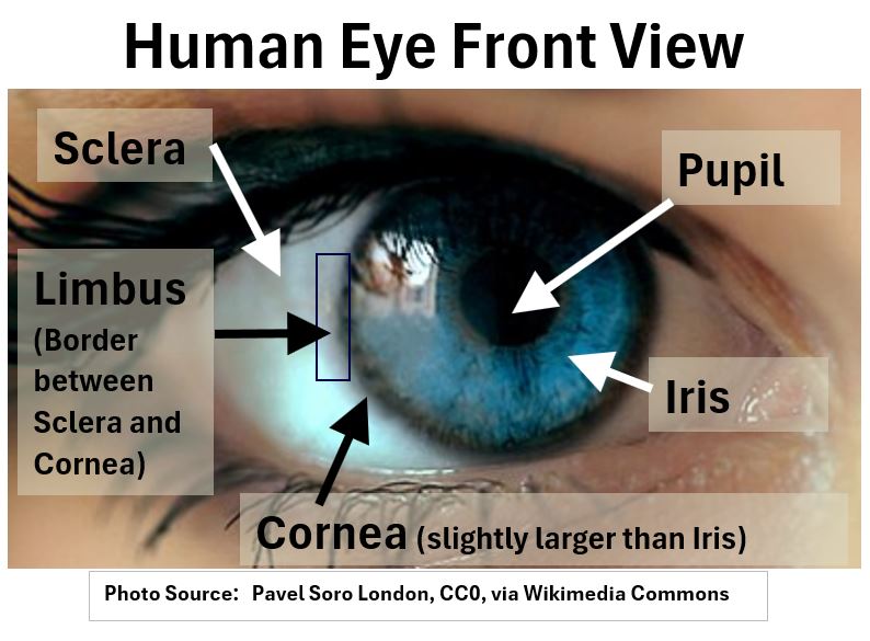
- the eyelid,
- the white of the eyeball (Sclera), which attaches to a
- domed shaped transparent Cornea, which covers a
- blue colored Iris and its Pupil (the central aperture or opening)
Picture_Human Eye Front View
Let’s take a more meaningful look using the cross sectional drawings of the eye below.
Picture_Human Eye Cross Section (Sagittal View)
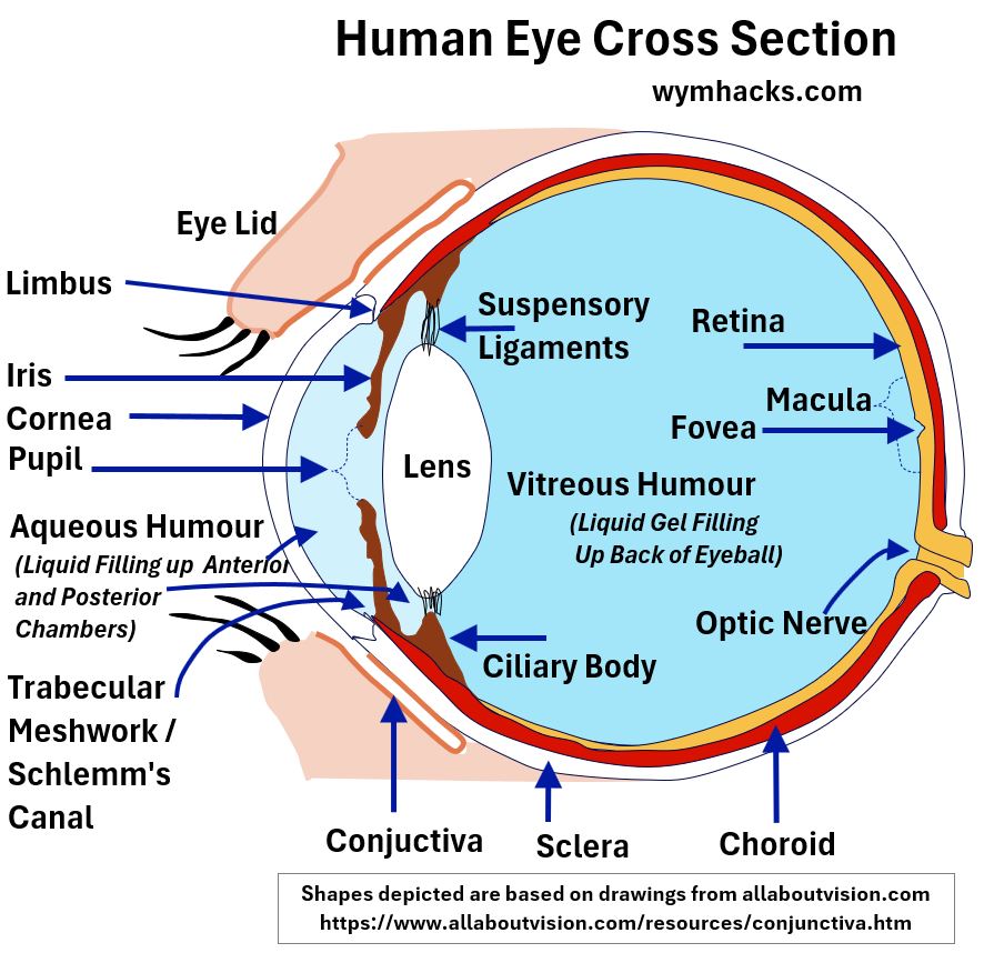
Note that the “Suspensory Ligaments” shown above are also known as Zonular Fibers or the Zonules of Zinn (named after an 18th century German anatomist).
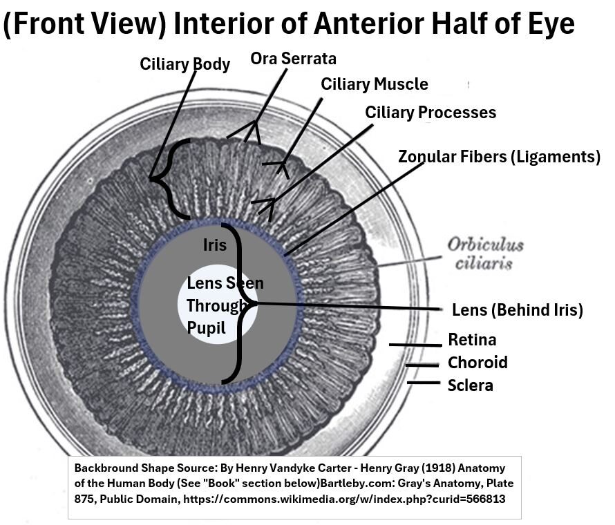
Get a little familiar with the internal structures in the schematic above, and then check out the “interior” sketch of a frontal view below.
This view is a good reminder that the components of the eye connect in a circular in nature.
Picture_Front View of Eye (Interior of Anterior Half)
Let’s start at the front (anterior) part of the eyes and identify/describe its major components.
Please refer to the above schematics as you read below to make sure you have a good dimensional feel for the eye’s structure.
Cornea
The cornea is that top domed “dark blue shaded” section in the drawing below.
It eventually transitions continuously into the Sclera through a transition zone called the Limbus.
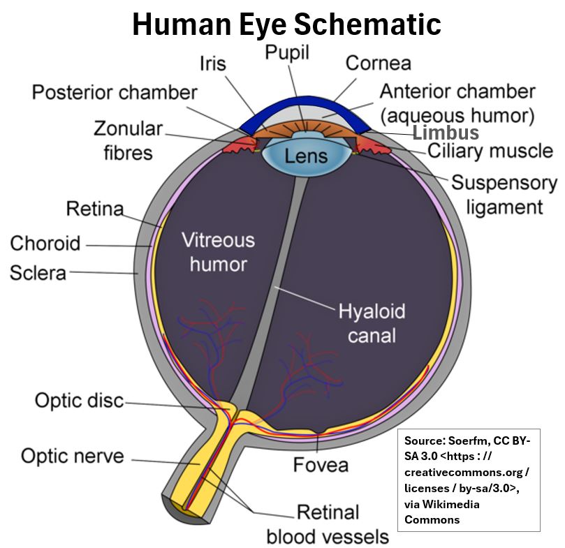
The word Cornea has a Latin etymology (meaning “horn”) and it’s hard for me to imagine the intended analogy.
I suppose you could imagine the cornea to be similar in shape to a symmetrical dome shaped horn.
- The Cornea is located in the outer central surface of the eye.
- It is dome shaped and transparent and covers the Iris and Lens beneath it.
- Its convex shape allows it to bend (refract) light and project (focus) it as an image on the back of your eyeball (Retina).
- This allows you to see of course.
- The Cornea provides about 2/3 of the total focusing power of your eye (the other 1/3 coming from the Lens).
- It also provides a protective layer against foreign objects (dust and other)
- It is 300 to 600 times as sensitive as skin and this trait allows it to trigger reflexes like blinking and tearing.
- The Cornea also has a multilayered tear film that keeps it moist and protected from infection.
This invisible light bending and protective layer is a critical component of your eye.
Although the cornea transitions into the Sclera, it is distinctly different in structure and function.
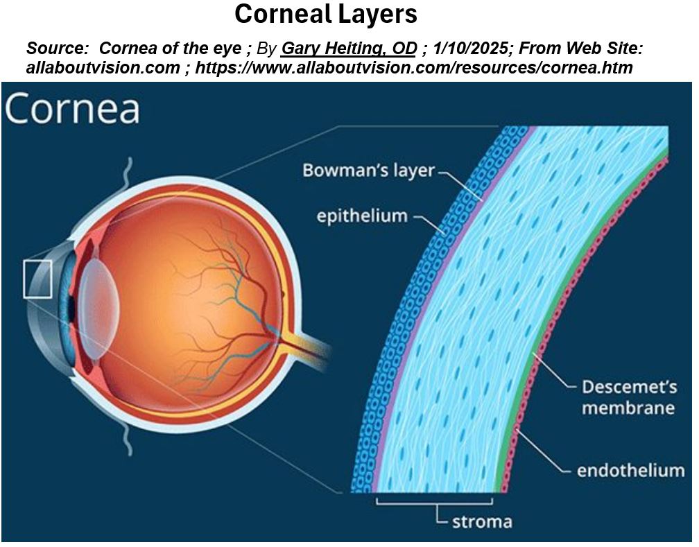
Corneal Layers
We see that the cornea is the major focusing component of the eyeball.
- Its shape (curvature) greatly influences our ability to see clearly.
- An irregularly shaped eye (an Astigmatic eye), which is rather common, can cause vision error, but
- there are surgical techniques to correct this.
Understanding the layers of the cornea will come in useful if you are exploring surgical solutions for correcting Astigmatism.
Picture_Corneal Layers
1. Epithelium Layer:
- Top (Surface) Layer about 50 microns (micrometers) thick (5 – 7 cell layers).
- Barrier to Water, Chemicals, and Microbes.
- Provides a smooth surface for tear film.
- Contains cells which provide immune protection (Langerhans Cells)
2. Bowman’s Layer:
- Located under Epithelium.
- A tough 8-12 micron thick layer that keeps the cornea from swelling forward
- Helps maintain the shape of the cornea
3. Stroma:
- The Stroma makes up about 90% of the Cornea.
- At about 500 microns thick, it provides mechanical strength.
- Comprises highly arranged collagen fibers and supporting keratocytes (enabling transparency)
- It’s the main refracting lens of the eye.
4. Descemet’s Layer:
- About 7 microns thick.
- Important for the health of endothelial cells.
- Major need for cornea transplant caused by damage to Descemet’s layer (Fuch’s dystrophy).
5. Endothelium:
- Works as a barrier and a pump. Removes water from the Stroma (keeping cornea clear).
Corneal layers information mostly sourced from:
Sclera
The Sclera (from the Greek word for hard) is a white colored, strong, fibrous tissue.
It wraps around the eye from the Cornea in the front to the Optic Nerve in the back.

- The border between the Sclera and the Cornea is called the Limbus.
- The Sclera helps maintain the eye’s shape and
- provides a layer of protection,
Limbus
The Limbus (Latin for “border”) is the circular transition area (1 – 2 millimeters wide) between the transparent Cornea and the white colored Sclera.


In Cataract removal surgery, a circumferential incision (cut) is typically made at the outer edge of the cornea, near the limbus (called a Clear Corneal Incision) .
The surgeon uses this opening to remove the bad lens and replace it with an artificial interocular lens (IOL).
Watch this fantastic Johns Hopkins animation of a small-incision cataract surgery (Phacoemulsification).
This video shows a manual surgery but laser technology can also be used to complete some of these steps.
Aqueous Humor
Assume that the spelling of Humor might differ depending on where you are (humor or humour).
Aqueous Humor is liquid (nutrients mixed in 99.9% water) that provides
- nourishment to the surrounding tissue and
- maintains the pressure of the eye (interocular) and therefore its shape integrity.

The Ciliary Body in a healthy eye continuously produces it and the Trabecular Meshwork and Schlemm’s Canal continuously drain it.
From there, the liquid is collected and routed to the Circulatory System.
- So, the direction of flow is “outwards” from the Posterior Chamber to the Anterior Chamber of the eyes.
- Too much production and/or too little drainage can cause high ocular pressure, resulting in Glaucoma.
If you want to learn more about this , check out these two articles which provide nice animations:
Iris / Ciliary Body / Choroid
The middle layer of the eye is called the Uvea (Latin for “grape”) because the “eye looks like a reddish-blue grape when the outer coat has been dissected away“.
The Uvea comprises the Iris , Ciliary Body, and Choroid.

Iris
Depending on the brightness of light, the Iris controls the size of its Pupil (the hole in the middle of it).
It’s a thin muscular structure that contains the pigment Melanin which gives your eye its color (Latin/Greek for rainbow).
The more Melanin, the darker your eyes (brown or darker).
Choroid
The Choroid layer is located between the Sclera and the Retina.
It’s a highly vascular membrane that provides nutrients and oxygen to the Retina.
Its etymology is from the Greek word for membrane.
Ciliary Body
The Ciliary Body surrounds the lens and keeps it in place (see the Interior Half of Anterior picture above).
The term “Ciliary” comes from the Latin word for “eyelash” as it resembles a fringe of eyelashes.
The Ciliary Body is a ring of muscle located behind the iris. It
- holds the Lens in place and it controls the focusing ability of the eye by changing the shape of the Lens.
- is connected to the Lens via a network of tiny ligaments called Suspensory Ligaments (or Zonular Fibers or Ciliary Zonules or Zonules of Zinn).
- continuously secretes Aqueous Humor, which fills the Posterior and Anterior Chambers and drains out via the Trabecular Meshwork.
Conjuctiva
The Conjuctiva is your inner eyelid membrane that connects to the front part of your eye. It
- lubricates the eye and inner eyelids,
- keeps irritants out, and
- with the tear glands, prevents infections.

The Conjuctiva makes mucus which combines with aqueous liquid (from Lacrimal Glands) and oily material (from Meibomian Glands) to make tears.
Tears contain enzymes that help to kill bacteria and viruses, flushing away potential pathogens.
Lens
The Lens is biconvex shaped (like a lentil!) and transparent.

- It’s composed of a crystalline compact structure, which minimizes light scattering and maintains transparency.
The Lens has several functions:
- It provides about 30% of the refractive power of the eye (the ability to bend/refract/converge incoming light into a focused and clear image on the Retina, at the Fovea).
- The Cornea provides about 70% of the refraction.
- The Lens performs fine tuned focusing by changing its shape (a process called Accomodation).
- It protects the Retina by absorbing UV radiation (Absorbs all UVB and most of UVA radiation). The Cornea also absorbs some UV radiation.
See my post on Eye Lens Anatomy for more details.
Vitreous Humor
The Vitreous Chamber of the eye is located behind (posterior to) the Lens.

This chamber makes up about 80% of the eyeball volume and is filled with a gelatinous liquid called Vitreous Humor (from the Latin, “glass-like”).
Vitreous Humor comprises mostly water , just like Aqueous Humor, but it contains chemicals that make it gelatinous (collagen, hyaluronic acid).
Vitreous Humor
- gives the eye its spherical structure.
- stays clear to allow for light to travel to the back of the eyeball.
- is a safety device, acting as a shock absorber.
Hyaloid Canal
The hyaloid canal (AKA Cloquet’s canal or Stilling’s canal):
- is a small channel that runs through the vitreous body of the eye.
- extends from the “optic nerve entrance to the eye” to the back of (posterior) of the lens.
- contains the fetal hyaloid artery which supplies nutrients to the developing lens.
- this artery is a fetal remnant i.e it is not active and has no significant function in adults (it typically “regresses”).
Retina
In each eye, the large chamber (~80% of volume of eye) behind the Lens is inflated with Vitreous Humor.
The inner layer of this chamber , called the Retina, contains photoreceptors and other nerve cells which take light rays from outside our eyes and convert them to electrical signals.

The electrical signals are routed via the Optic Nerve to various areas of the brain which translates these into vision and color.
The Eye is just an extension of the brain, located at the surface, like the periscope of a submarine.
Picture_The Eye is an Extension of the Brain
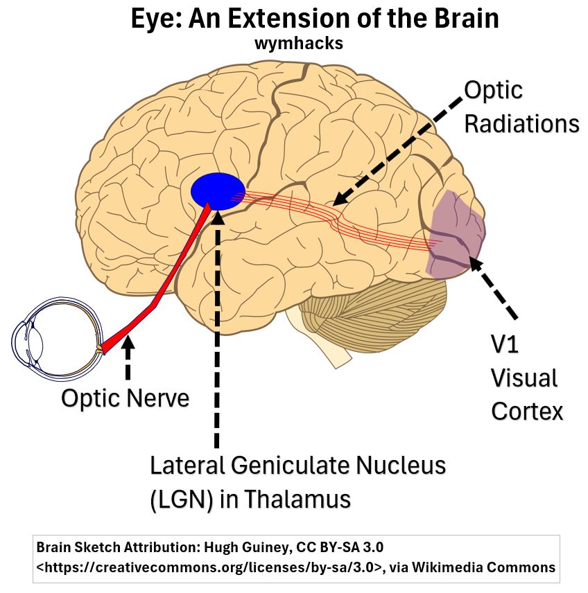
If you want to learn more about vision and color check out my article: A Color Primer (Including How to Create an Effective Colour Palette)
The word Retina comes from the Latin for “net” (like a fishing net).
I think this is in reference to the eye blood vessels i.e. Imagine the blood vessels coming in via the Optic Nerve of the eye and then branching out across the eyeball like a net.
The Muslim Scholar Ibn Ishak (860 AD) used the Arabic word Shabakeh (net) to describe the Retina and this was eventually translated back into Latin and English.
- You should know that during the Muslim era (8th – 13th centuries), scholars preserved many ancient Greek and Latin scientific learnings by translating them into Arabic.
- These were ultimately translated back into our current modern idioms.
- There were many original scientific works published as well, especially regarding the eye and optics.
In the picture below, I show an eye zoom-out of a Macular section of the Retina and another zoom-out showing the photoreceptor Rod and Cone Cells.
The Macula is a region that has the highest counts of photoreceptor cells and is where your eyes focus to give you clear and colorful vision.
In the center of the Macula is an indented area called the Fovea which contains the highest density of color sensing Cone photoreceptors (and no Rod Cells) .
Cone Cells sense color and provide vision clarity (acuity or sharpness).
Picture_Retinal Layers and Rods and Cone Photoreceptors
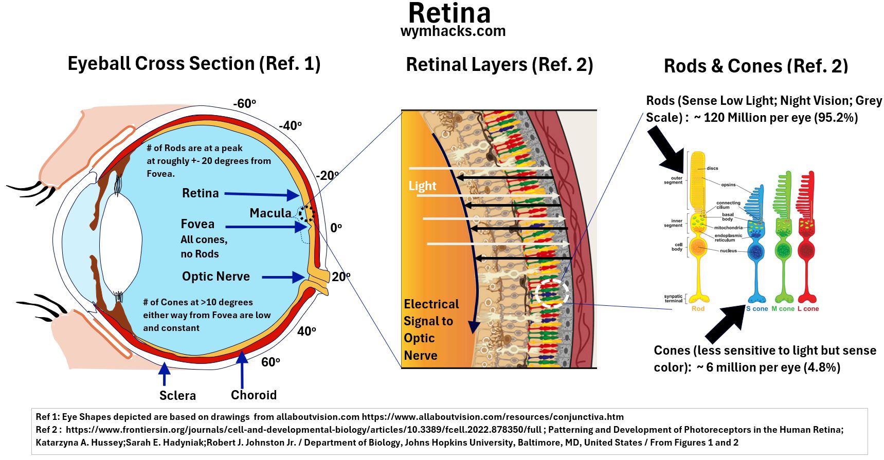
There is a lot to digest in the picture above, and it’s probably hard to see if you are using a hand held device.
So I’ll break the picture down into larger sections below.
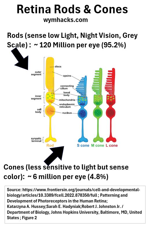
Let’s explore this drawing going from right to left starting with the Rod and Cone Photoreceptors. Look at a close up of these cells below.
Picture_Rod and Cone Photoreceptors
According to aao.org, the Retina contains approximately 126 million Photoreceptor cells of which
- 120 million (95.2%) are Rod Cells and
- 6 million (4.8%) are Cone Cells.
Rods operate at low levels of illumination (scotopic environment) and helps us see at night in shades of Grey.
As illumination increases (photopic environment) , Rods are no longer in play and the Cones now take over.
Cone Cells are less sensitive to light than Rod Cells but can sense color. There are three types of Cones Cells:
- S or Blue Cones (S = short wavelength light i.e. most sensitive to Blue Light)
- M or Green Cones (M = medium wavelength light i.e. most sensitive Green Light)
- L or Red Cones (L = long wavelength light i.e. most sensitive to Red Light)
Even though these are described with a specific color, they actually sense a range of colors but are most sensitive to their eponymous color (their namesake color).
See my Color Primer article for more on this.
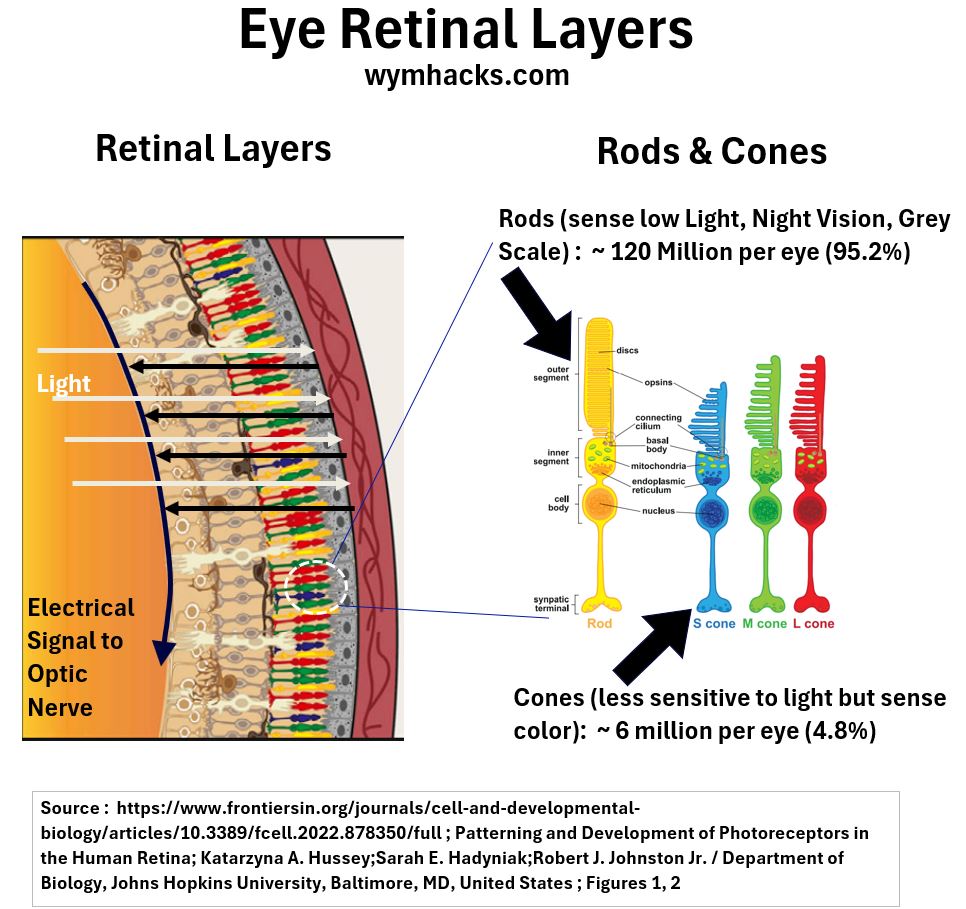
Ok, so how are these photoreceptors situated and how does light become sight?
Picture_Retinal Layers and Paths of Light and Electrical Impulses
Here is a very high level description of the visual pathway:
- Light (the white lines in the drawing), seemingly non-intuitively, travels several layers into the Retina and is absorbed by a cell layer called the Retinal Pigment Epithelium or RPE (perhaps to regenerate pigment producing layers in this area).
- Photoreceptor Cells (Rod and Cone Cells), one layer up from the RPE, receive the light and convert it into electrical signals using a process called Phototransduction.
- In Phototransduction light energy changes the chemical structure of a pigment called Retinal, which causes a change in electrical potential.
- The electrical signals now travel the opposite way back up the nerve cell layers of the Retina (the black lines in the drawing).
- These electrical signals , relayed and regulated by various nerve cells (Horizontal Cells, Bipolar Cells, Amacrine Cells, Retinal Ganglion Cells), are ultimately routed down an electrical nerve “highway” called the Optic Nerve (via Neuron cells).
- The Optic Nerve relays the signals to the brain (to Thalamus and Visual Cortex) which make the pictures we see (wild).
The below are two good references if you want to learn more:
The whole of the Retina, the inner layer of the eyeball in the picture below, contains Photoreceptors, ~ 95.2% of which are Rod Cells.
The next two drawings below show how these photoreceptors are distributed.
Picture_Retina Showing Degrees Angle from Fovea
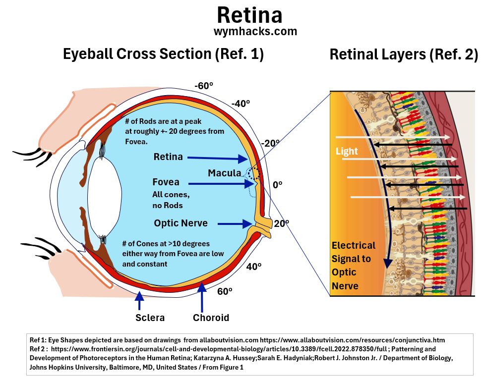
Notice the degree angles that are noted around the eyeball; with the 0 degree angle marking the location of the Fovea.
Recall that the Fovea is located at the center of the Macula and contains the highest density of Cone Cells (and no Rod Cells).
In the picture above, the zoom-out of a Macular region shows a large number of Cone Cells and a few Rod Cells.
The Fovea however will only contain Cone Cells.
The graph below plots the density of the Rod and Cone Cells relative to their distance from the Fovea (located at angle 0 degrees)
Picture_Rod and Cone Cell Retinal Distribution
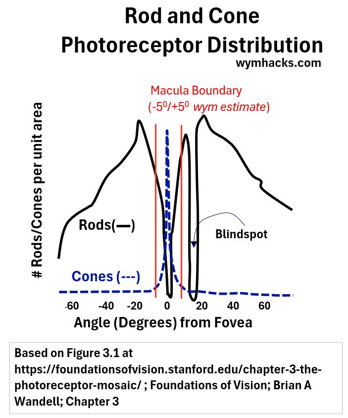
The above graph
- shows that for most of the Retinal surface, Cone Cell quantities (blue dotted line) are low and constant (relative to the much more plentiful Rod Cells).
- shows how the Cone Cell density spikes at the Fovea , the center of the Macula.
So, the Fovea is where our eyes need to focus to maximize color perception and sharpness of vision.
Conclusion
The human eye is a complex organ that functions as an extension of the brain, translating light into color and vision.
It receives light rays from the environment and primarily refracts them through the cornea and the lens, projecting them onto the retina.
These light signals are then converted into electrical impulses by specialized cells in the retina.
These electrical signals are transmitted to the brain via the optic nerve, where they are processed and interpreted as the images we ‘see’.
Disclaimer: The content of this article is intended for general informational and recreational purposes only and is not a substitute for professional “advice”. We are not responsible for your decisions and actions. Refer to our Disclaimer Page.
