Menu (linked Index)
Eye Lens Anatomy and Accommodation
Last Update: December 22, 2024
Introduction
After my cataract surgery in 2024, I decided to write an article on the experience.
The effort spawned a few more articles on the anatomy and physics of the eye.
This article describes the anatomy of the Lens; that
- transparent (at least begins that way)
- refracting piece of your eye that
- helps direct light to your Retina.
After giving you a little description of the anatomy, I’ll describe generally how lenses are removed during Cataract Surgery.
I also describe your eye’s ability to focus light by changing the shape of your lens (called Accommodation).
Find more links related to my eye research here: Understand Your Eyes.
Lens Anatomy
The eye Lens gets its name from the Latin word for “Lentil” which is a biconvex shaped legume. Lentils have an interesting ancient history described nicely in this NPR article.
While we are there, the Greek word for lentil is “fakos”.
- “Phakic” refers to the natural eye Lens”
- “Pseudo-Phakic” refers to artificial Lens that have replaced the natural Lens.
- “Phakic emulsification or Phacoemulsification” is a surgical technique used to remove the natural Lens of the eye, often as part of cataract surgery.
Picture_Lentils

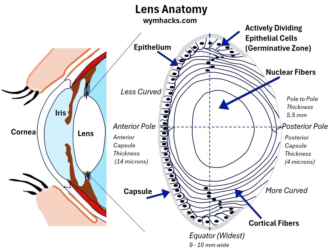
Consider the sagittal (vertical cross sectional) picture of an eye below.
I show the lens in relation to the eye, and, just to make sure you see its anatomy adequately, I also show a larger picture of it “rolled” onto its Posterior pole.
The Lens is biconvex shaped (like a lentil!) and transparent.
- It’s composed of a crystalline compact structure, which minimizes light scattering and maintains transparency.
The Lens has several functions:
- It provides about 30% of the refractive power of the eye (the ability to bend/refract/converge incoming light into a focused and clear image on the Retina, at the Fovea).
- The Cornea provides about 70% of the refraction.
- The Lens performs fine tuned focusing by changing its shape (a process called Accomodation).
- It protects the Retina by absorbing UV radiation (Absorbs all UVB and most of UVA radiation). The Cornea also absorbs some UV radiation.
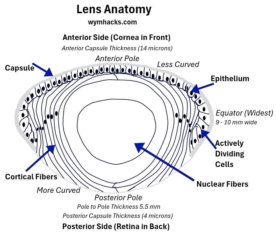
Picture_Lens Anatomy (Side View)


Lens Dimensions:
- The Anterior (less curved) and Posterior surfaces (more curved) meet at the Equator.
- Equator diameter is about 9 – 10 mm in an adult’s lens
- The thickness (from anterior to Posterior Pole) is about 5.5 mm
- The Anterior Pole is about 3mm distance from the Cornea
Lens Capsule:
The outer layer of the Lens is called the Capsule.
- This elastic membrane is thickest at the equatorial ends and thinnest at the Posterior Pole.
- In Cataract removal surgery a circular cut is made into the Anterior Capsule (called a Capsulorhexis or Anterior Capsulotomy).
- In Cataract removal surgery, a potential complication is damaging (rupturing) the Posterior Capsule.
Lens Epithelium:
The next layer, which does not encompass the whole Lens, is the Epithelium.
- The Lens Epithelium are “live” cells located on the Anterior surface and Equatorial ends.
- They are cuboidal on the Anterior side and are metabolically active.
- The Equatorial Epithelium cells divide and elongate into fibers which move to the posterior and interior.
Lens Cortex and Nucleus:
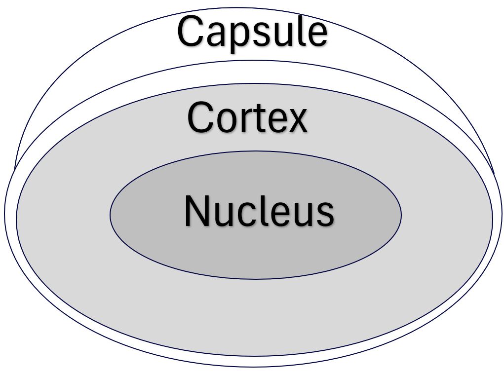
- The Epithelial cell growth continues forming onion like fiber layers. The newer outer layers represent the Cortex region.
- The Nucleus is the center area of the Lens and is the oldest and most dense section.
- The Cortex (Cortical Fibers) and Nucleus (Nuclear Fibers) are removed during Cataract removal surgery.
Cataract Surgery Impact on the Lens
In Cataract removal surgery, the Cortex and Nucleus are broken up and removed and replaced with an artificial lens (Interocular lens).
In order to perform this natural lens replacement , the surgeon has to make a circular cut into the Anterior Capsule (called a Capsulorhexis or Anterior Capsulotomy).
There are some potential complications and side effects of this surgery:
- A potential complication is damaging (rupturing) the Posterior Capsule (remember it’s thin on the posterior side). Vitreous Humor leakage can then occur.
- Another possibility, post surgery, is that some of the remaining Capsule will itself develop Cataracts (called a Secondary Cataract or Posterior Capsular Opacification or PCO)
- Caused by epithelial cells migrating to the posterior side.
- This is very common and is correctable with additional laser surgery
Lens Accommodation
The Lens , as we noted, provides about 30% of the refractive power of the eyes (the ability to bend light so it can be transformed into the image of an object by our Retina and Brain).
But, it also provides fine tuning functionality by changing the shape of the Lens. This process is called Accommodation.
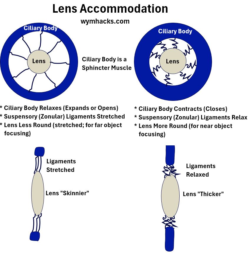
Check out the drawing below that shows anterior (front) and side views of the lens and surrounding “attachments”:
- The Ciliary Body is a sphincter muscle that can relax (expand) and contract (restrict).
- This will respectively stretch or relax the Suspensory Ligaments (the Zonules) which
- respectively stretches (to see a clear distant image) or thickens (to see a clear close image) the Lens.
Picture_Lens Accommodation (Anterior and Side Views)

This is kind of gross, but, the ciliary bodies act like their fellow sphincter muscle, your anus.
- Normally, in the relaxed state, your anus will look like the picture on the left (above) and
- ,when you are doing your “business”, you will have occasion to contract it, as in the picture (above) on the right.
Often models of the eye are provided and certain descriptors (like eye “power” or ability to focus) are provided based on the eye being in a “relaxed condition“.
This specifically means the Ciliary Bodies are in a relaxed state which means the eye Lens is at its maximum length (the left picture above).
Presbyopia (Loss of Accommodation)
As we age, the lens age and become more rigid.
- In an eventually universal eye condition called Presbyopia, the lens loses its flexibility and becomes less able to change shape to accommodate for different distances.
- This mainly results in difficulty focusing on nearby objects.
- Presbyopia eventually happens to everyone as they age and can be corrected via eyeglasses, contact lenses, surgeries or Eye drops.
Cataract Surgery Means No Accommodation
This amazing but sadly youthful Accommodation ability is completely lost when the natural lens is removed during Cataract surgery.
The ability to see near is then provided by the new artificial lens and/or reading glasses.
Conclusion
The lens, a transparent and flexible focusing device, directs rays of light to the back of the eye.
The eye manipulates the lens to fine-focus light, a process called Accommodation.
The Lens comprises multiple layers, from the outside in: the Capsule, the Epithelium, the Cortex, and the Nucleus.
During Cataract surgery, the lens is removed by making an incision in the capsule and then removing the internal contents (cortex and nucleus).
Accommodation ability weakens with age (Presbyopia) and Cataract surgery permanently eliminates it.
Disclaimer: The content of this article is intended for general informational and recreational purposes only and is not a substitute for professional “advice”. We are not responsible for your decisions and actions. Refer to our Disclaimer Page.

